ISIC 2018: Skin Lesion Analysis Towards Melanoma Detection - Task 3

Task 3: Lesion Diagnosis
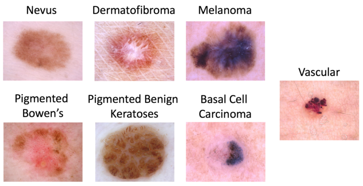
Capitation example

Aim

Submit automated predictions of disease classification within dermoscopic images. Possible disease categories are:

  1. Melanoma
  2. Melanocytic nevus
  3. Basal cell carcinoma
  4. Actinic keratosis / Bowen’s disease (intraepithelial carcinoma)
  5. Benign keratosis (solar lentigo / seborrheic keratosis / lichen planus-like keratosis)
  6. Dermatofibroma
  7. Vascular lesion

Background

Skin cancer is the most common cancer globally, with melanoma being the most deadly form. Dermoscopy is a skin imaging modality that has demonstrated improvement for diagnosis of skin cancer compared to unaided visual inspection. However, clinicians should receive adequate training for those improvements to be realized. In order to make expertise more widely available, the International Skin Imaging Collaboration (ISIC) has developed the ISIC Archive, an international repository of dermoscopic images, for both the purposes of clinical training, and for supporting technical research toward automated algorithmic analysis by hosting the ISIC Challenges.

Results

alt text

Challenge

https://challenge2018.isic-archive.com

Nils Gessert
Nils Gessert
Research Scientist
Thilo Sentker
Thilo Sentker
Research Scientist
Frederic Madesta
Frederic Madesta
PhD Student
Rüdiger Schmitz
Rüdiger Schmitz
PhD Student
Helge Kniep
Helge Kniep
PhD Student
Ivo Matteo Baltruschat
Co-Founder / Research Scientist

-Ivo Matteo Baltruschat studied Information and Electrical Engineering at the University of Applied Sciences, Hamburg between 2010 and 2014. In 2016, he finished his Master of Science in medical engineering science at the Universität zu Lübeck with his thesis on Deep Learning for Advanced Medical Applications. Currently, he is a PhD student in the group of Tobias Knopp at the Institute for Biomedical Imaging, Hamburg University of Technology. His research covers the automatic analysis of medical x-ray images using machine learning methods. Our objective is to apply deep learning methods to high-resolution medical x-ray images. In the long-term, we are pursuing the goal to employ deep learning methods in order to find patterns between medical reports and medical x-ray images that will help to push the state-of-the-art in computer-aided diagnosis for chest x-ray images.

René Werner
René Werner
Co-Founder / Head of scientific working group