StructSeg 2019 - Task 3
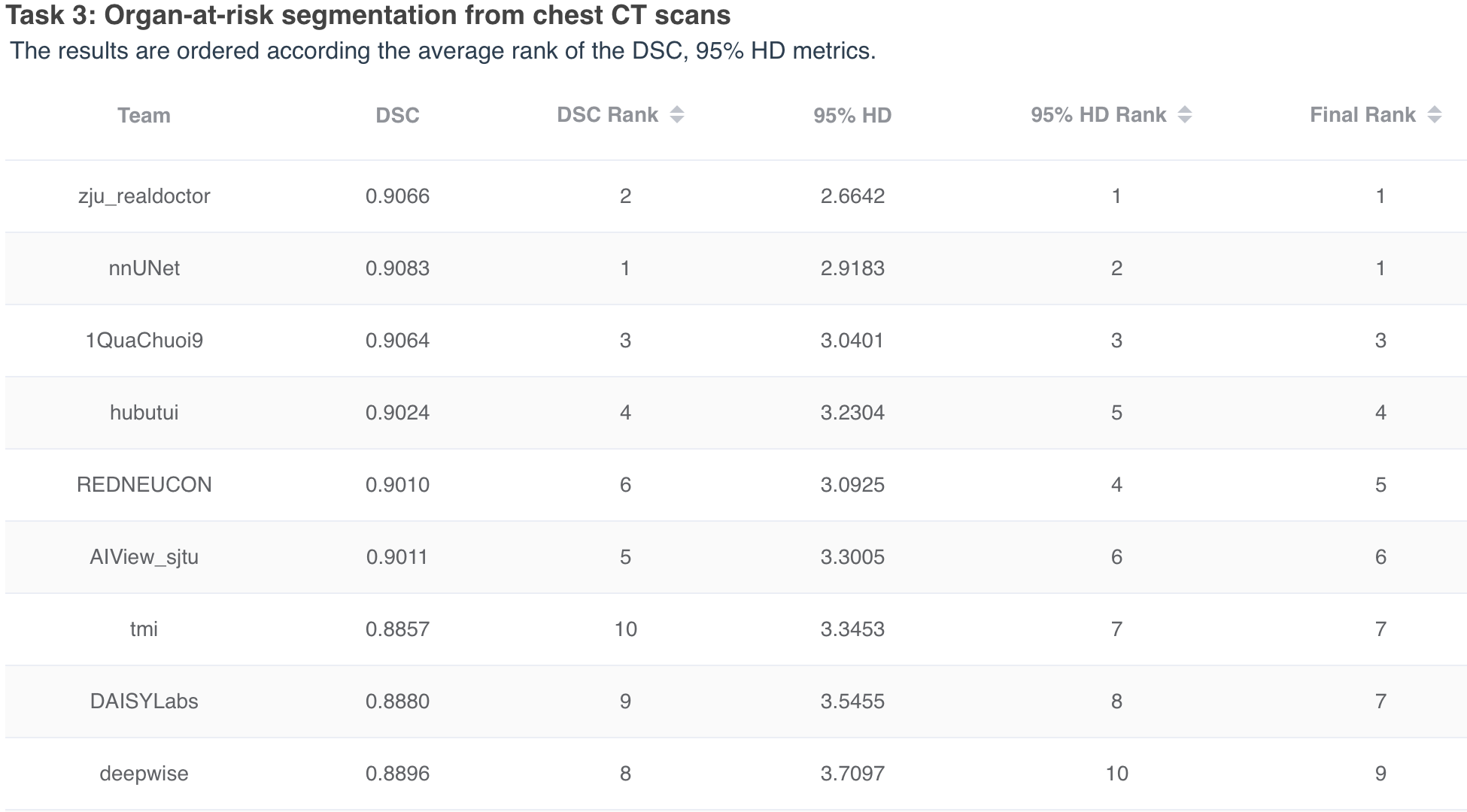
Task 3: Organ-at-risk segmentation from chest CT scans
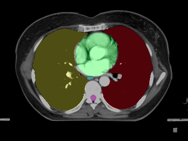 Capitation example
Aim
The goal of this task was the segmentation of six organ-at-risk in chest CT scans:
- left lung
- right lung
- spinal cord
- esophagus
- heart
- trachea
Background
Radiation therapy is one type of important cancer treatment for killing cancer cells with external beam radiation. Treatment planning is vital for the treatment, which sets up the radiation dose distribution for tumor and ordinary organs. The goal of planning is to ensure the cancer cells receiving enough radiation and to prevent normal cells in organs-at-risk (OAR) from being damaged too much. Organs-at-risk are usually the organs that are sensitive to radiation. For instance, optical nerves and chiasma in the head cannot receive too much radiation otherwise the patient risks losing his/her vision. Gross Target Volume (GTV) is the position and extent of gross tumor imaged by CT scans, i.e. what can be seen.
One important step in radiotherapy treatment planning is therefore to delineate the boundaries of tens of OARs and GTV in every slice of a patient’s CT scans, which is tedious and occupies much of oncologists’ time. Automatic OAR and GTV delineation would substantially reduce the treatment planning time and therefore reduce the overall cost for radiotherapy.
Results