Aim
The goal for ISIC 2019: Task 1 is classify dermoscopic images without meta-data among nine different diagnostic categories:
- Melanoma
- Melanocytic nevus
- Basal cell carcinoma
- Actinic keratosis
- Benign keratosis (solar lentigo / seborrheic keratosis / lichen planus-like keratosis)
- Dermatofibroma
- Vascular lesion
- Squamous cell carcinoma
- None of the others
Background
Skin cancer is a major public health problem, with over 5,000,000 newly diagnosed cases in the United States every year. Melanoma is the deadliest form of skin cancer, responsible for an overwhelming majority of skin cancer deaths. In 2015, the global incidence of melanoma was estimated to be over 350,000 cases, with almost 60,000 deaths. Although the mortality is significant, when detected early, melanoma survival exceeds 95%.
About Dermoscopy
As pigmented lesions occurring on the surface of the skin, melanoma is amenable to early detection by expert visual inspection. It is also amenable to automated detection with image analysis. Given the widespread availability of high-resolution cameras, algorithms that can improve our ability to screen and detect troublesome lesions can be of great value. As a result, many centers have begun their own research efforts on automated analysis. However, a centralized, coordinated, and comparative effort across institutions has yet to be implemented. Dermoscopy is an imaging technique that eliminates the surface reflection of skin. By removing surface reflection, visualization of deeper levels of skin is enhanced. Prior research has shown that when used by expert dermatologists, dermoscopy provides improved diagnostic accuracy, in comparison to standard photography. As inexpensive consumer dermatoscope attachments for smart phones are beginning to reach the market, the opportunity for automated dermoscopic assessment algorithms to positively influence patient care increases.
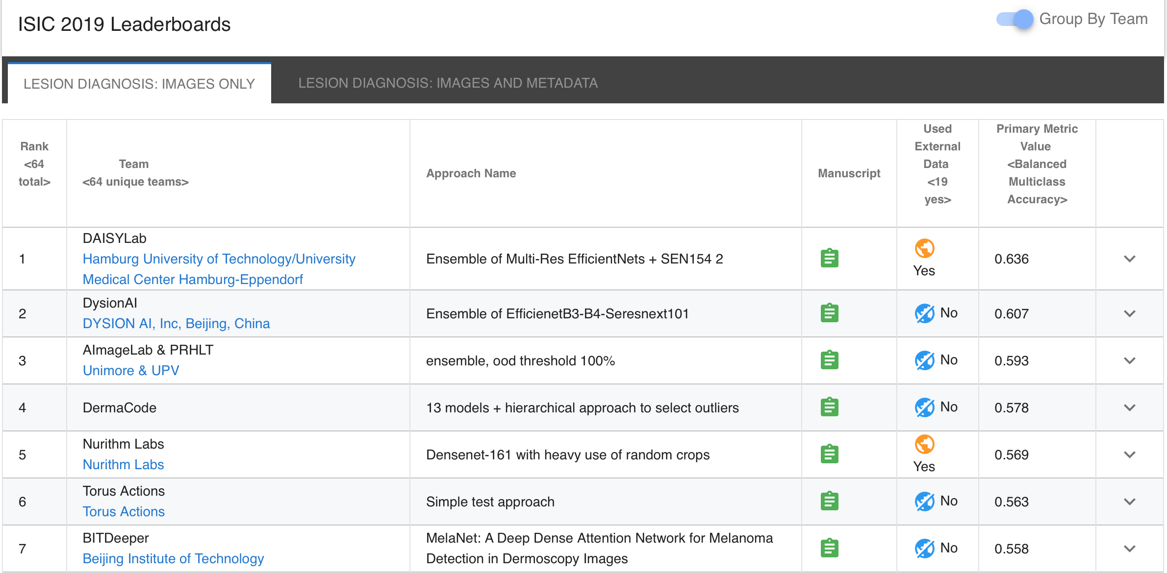
Results
Challenge
Nils Gessert
Research Scientist
Maximilian Nielsen
Student
Mohsin Shaikh
MSc Student
Ivo Matteo Baltruschat
Co-Founder / Research Scientist
Ivo Matteo Baltruschat studied Information and Electrical Engineering at the University of Applied Sciences, Hamburg between 2010 and 2014. In 2016, he finished his Master of Science in medical engineering science at the Universität zu Lübeck with his thesis on Deep Learning for Advanced Medical Applications. During DAISYlabs, he was a PhD student in the group of Tobias Knopp at the Institute for Biomedical Imaging, Hamburg University of Technology. His research covered the automatic analysis of medical x-ray images using machine learning methods, with a focus on applying deep learning to high-resolution medical x-ray images for computer-aided diagnosis.
Rüdiger Schmitz
PhD Student
René Werner
Co-Founder / Head of scientific working group
René Werner co-founded DAISYlabs and led the scientific working group at the University Medical Center Hamburg-Eppendorf, coordinating deep learning research for biomedical image processing across UKE and TUHH.